
Table of Contents
Very Small Objects Have Big Impact on Health
International workshop focuses on nanoscale modeling and imaging of biological systems
Alzheimer’s disease, cystic fibrosis, Parkinson’s disease and Type 2 diabetes are among many inherited diseases that hold their secrets at the nanoscale level.
Unlocking these secrets is challenging, requiring the study of nanoscale biological objects, whose size is calculated in billionths of a meter. For comparison, the width of a human hair is approximately 80,000 nanometers. One inch is approximately 25 million nanometers.
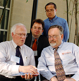
Nanoscale modeling workshop keynote speaker Robert
M.
Glaeser, Ph.D., left, talks with, from left, workshop
organizer
Willy Wriggers, Ph.D.; presenter Zhiyong
Zhang, Ph.D.,
fellow at the UT School of Health
Information Sciences at
Houston (SHIS); and SHIS
Dean Jack Smith, M.D., Ph.D.
Photo by Ester Fant
“Researchers are now able to use advanced electron microscopic imaging, x-ray crystallography, bioinformatics and computer modeling of nanoscale biological objects and events to explore normal and disease processes,” said Jack Smith, M.D., Ph.D., dean of The University of Texas School of Health Information Sciences at Houston (SHIS). “Such computer modeling can significantly speed up our understanding of these phenomena over traditional laboratory experiments.”
A six-day international workshop in late April on “Innovations in Nanoscale Modeling and Imaging of Biological Systems” brought to the Texas Medical Center world leaders in nanoscale biological system modeling to share their knowledge and teach others to use their methods and software systems. A second goal was to foster an alliance with innovative researchers in the private sector and academia so software developers at SHIS can build on their strengths in a compatible mode with leading laboratories around the world.
The workshop featured 21 invited speakers and 50 invited participants from as far away as Germany, Israel, Singapore, South Africa, Spain and the United Kingdom. In addition, about 30 Houston-area scientists attended the public lectures.
Hands-on sessions trained participants from the fields of electron microscopy and x-ray diffraction in the use of modeling, imaging and visualization software developed by selected speakers and by the laboratory of conference chairman Willy Wriggers, Ph.D., assistant professor at SHIS. The sessions emphasized interfaces with compatible software used in structural biology.
“These were mainly junior faculty at other institutions whom I know from my research and from international conferences,” said Wriggers, who also holds a faculty appointment at the Brown Foundation Institute of Molecular Medicine for the Prevention of Human Diseases. “I’ve had past collaborations with most of them so I knew their software works well with ours in a compatible mode.
“Many new collaborations were fostered during this time, and we were able to connect with the users of our software, who presented their own work on posters during the workshop,” Wriggers said. “We appreciate the publications that cite our methods, but it is an entirely new experience to meet with the enthusiastic authors in person. You get this great sense of appreciation and accomplishment directly from the community.”
Workshop participant Danny Doan, Ph.D., expects the software developed by Wriggers and his team to be valuable in solving the atomic structure of the p53 tumor suppressor protein. “Information on the p53 protein at the structural level can lead to success in designing anti-cancer drugs,” said Doan, who is a postdoctoral fellow at the Institute of Molecular and Cell Biology in Singapore.
Another participant, Oscar Llorca, Ph.D., said the meeting covered both the theoretical background and the practical, everyday applications of “state-of-the-art methodologies in the analysis and interpretation of structures derived from electron microscopy images.” At the Centre for Biological Research–Spanish National Research Council in Madrid, Llorca heads a research group that uses electron microscopy and 3D reconstruction in the structural analyses of large macromolecular complexes relevant to biomedicine.
The keynote speaker, Robert M. Glaeser, Ph.D., is a world-renowned electron microscopist who is professor in the Department of Molecular and Cell Biology and the Lawrence Berkeley National Laboratory at the University of California at Berkeley. “His keynote lecture showed that even with his seniority he still has another lifetime of ideas in him,” Wriggers said. “Many of us younger scientists in the audience were energized by his continuing enthusiasm for scientific progress.”
Computer scientist Bimal Rath, Ph.D., from the New York State Department of Health, was impressed by both the program and the “top of the line” facilities at SHIS. “The presenters were excellent and are well known in their respective fields for their cuttingedge research. The workshop was organized superbly and everything went very, very smoothly,” said Rath, who contributed to developing an image processing system that is being used in more than 100 universities and research institutions worldwide.
Participants expressed their appreciation to Wriggers and his coworkers, in particular postdoctoral fellows Jochen Heyd, Ph.D., and Stefan Birmanns, Ph.D., as well as administrative assistant Namiko Burleson and event planner Hilary Wriggers, for organizing the workshop.
Wriggers thanked the John P. McGovern Foundation and Tejas Office Products for their support.
By Ina Fried, Public Affairs
Next article